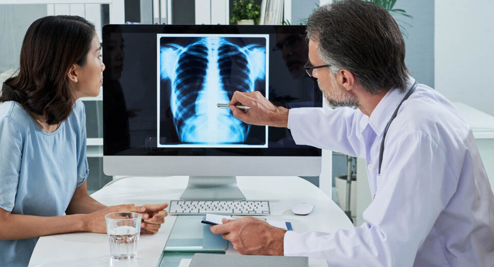
Imaging is one of the most powerful tools in modern medicine for the early detection of cancer. Unlike blood tests or physical exams, imaging allows providers to see what’s happening inside the body—often before any …
The Role of Imaging in Cancer Screening
July 12, 2025
Imaging is one of the most powerful tools in modern medicine for the early detection of cancer. Unlike blood tests or physical exams, imaging allows providers to see what’s happening inside the body—often before any symptoms are noticeable. These tests can help detect tumors in their earliest stages, when they are more likely to be treated successfully.
From mammograms to CT scans and MRIs, imaging supports both routine screening and the evaluation of specific concerns. Understanding how and when these tools are used gives patients greater confidence in their care and highlights the importance of early intervention.
Why imaging matters in early detection
Cancer often develops silently. In many cases, there are no outward signs until the disease has grown or spread. Imaging allows for visual identification of tumors, cysts, and structural abnormalities—sometimes long before they cause physical symptoms.
The earlier cancer is found, the greater the chance of successful treatment. Imaging helps identify changes in tissue or organ function, providing a foundation for diagnosis and guiding the next steps in care. It can also help determine the stage of a known cancer, which affects treatment decisions.
Common imaging tests used in screening
Different types of imaging are used depending on the organ being screened, the patient’s risk level, and the type of cancer being evaluated. Each test has its own strengths, preparation requirements, and limitations.
Mammogram
Used primarily for breast cancer screening, mammograms are low-dose X-rays that can detect tumors too small to be felt. They are recommended regularly for women over 40, depending on individual risk factors.
Low-dose CT scan (LDCT)
This test is used to screen for lung cancer in high-risk individuals, such as long-term smokers. It uses lower radiation than a standard CT and can detect nodules or masses in the lungs before symptoms develop.
Colonoscopy with imaging
While colonoscopy involves direct visualization rather than traditional imaging, CT colonography (also called virtual colonoscopy) uses imaging technology to screen the colon for growths and abnormalities without invasive instruments.
Ultrasound
Ultrasound is often used in abdominal, pelvic, and thyroid cancer screening. It uses sound waves to create images of internal organs and is helpful in identifying masses or fluid-filled cysts.
MRI (Magnetic Resonance Imaging)
MRI is often used when detailed soft-tissue imaging is needed, such as in the brain, spine, or breast. It can offer additional information after a mammogram or when screening for cancers in individuals with dense tissue or genetic risk factors.
PET scan (Positron Emission Tomography)
PET scans are more commonly used after diagnosis, but they play a role in staging and assessing the spread of certain cancers. These tests detect metabolic activity that may signal aggressive or advanced disease.
When imaging is recommended
Imaging is not always used for general screening in healthy individuals. Most imaging-based screenings are recommended based on risk factors like age, family history, smoking, or the presence of symptoms.
Some standard examples include:
- Mammograms every 1–2 years for women over 40
- Annual low-dose CT scans for adults aged 50–80 with a smoking history
- Ultrasounds for women with ovarian cancer risk factors
- Colonoscopy or CT colonography beginning at age 45 for colorectal cancer screening
For individuals with a personal or family history of certain cancers, earlier or more frequent imaging may be recommended.
Benefits and limitations of imaging tests
Imaging plays a critical role in finding cancer early, but it’s important to understand both its strengths and limitations.
Benefits include:
- Ability to detect cancers before symptoms appear
- Guidance for biopsies and follow-up testing
- Non-invasive or minimally invasive nature of many tests
- Monitoring disease progression or treatment response
Limitations include:
- False positives, which may lead to unnecessary follow-up tests
- False negatives, especially with small or slow-growing tumors
- Exposure to radiation in some tests, such as CT scans
- Variability in interpretation between radiologists
Despite these considerations, the benefits of imaging in high-risk or symptomatic individuals typically outweigh the drawbacks.
Imaging as part of a complete screening plan
While imaging is powerful, it is most effective when used alongside other screening tools like blood tests, physical exams, and family history evaluations. A full preventive care plan considers all available data points to catch disease as early as possible.
For example, a woman with a family history of breast cancer might receive mammograms and MRI screenings, while a man at high risk for colorectal cancer may undergo earlier imaging combined with stool-based tests.
Imaging also plays a role in post-treatment monitoring. After cancer has been treated, periodic imaging helps track recurrence or evaluate the response to therapy. These follow-ups can be vital in adjusting treatment plans and maintaining long-term health.
Takeaway
Imaging technologies have transformed the way cancer is screened and diagnosed. By making it possible to visualize abnormalities at an early stage, these tools offer patients and providers a chance to act sooner and more effectively. Whether part of a routine exam or a targeted investigation, imaging continues to be a cornerstone of preventive cancer care.