Medical imaging has evolved rapidly over the past two decades, and one of the most significant improvements has been the shift from traditional film-based X-rays to digital X-ray technology. This advancement offers more than just …
Digital X‑Rays: Faster Results, Lower Exposure
July 12, 2025
Medical imaging has evolved rapidly over the past two decades, and one of the most significant improvements has been the shift from traditional film-based X-rays to digital X-ray technology. This advancement offers more than just convenience—it delivers faster results, greater precision, and lower radiation exposure for patients.
If you’re scheduled for an imaging test or considering an urgent care visit for musculoskeletal or chest concerns, understanding the benefits of digital X-rays can help you feel more informed about your care.
What are digital X-rays?
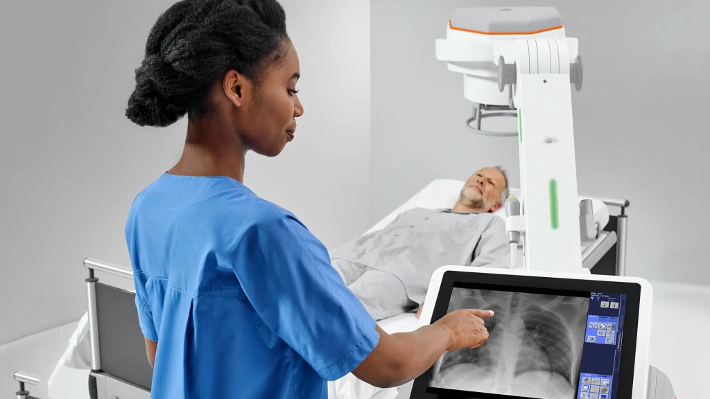
Digital X-rays use electronic sensors instead of traditional photographic film to capture images of the body’s internal structures. These sensors convert the X-ray energy into digital data, which is immediately processed and displayed on a computer screen.
Unlike traditional film X-rays, which require time for chemical development, digital X-rays produce images almost instantly—reducing wait times and allowing for faster diagnosis.
How digital X-rays improve patient experience
One of the biggest advantages of digital imaging is speed. In urgent care, primary care, or hospital settings, faster image turnaround can make a significant difference in both diagnosis and treatment.
Here’s how digital X-rays enhance the patient experience:
- Immediate viewing: Providers can see and analyze the images in real time
- Quicker diagnosis: Fast results help guide immediate decisions, especially for injuries or infections
- Less time in the exam room: Shorter exposure times and fewer repeat images mean a faster visit overall
- Better communication: Images can be shared with specialists quickly for consultation
The efficiency of digital imaging also improves clinical workflows, which means shorter wait times for patients and more coordinated care.
Lower radiation exposure with digital imaging
Radiation safety is a concern for many patients, especially for those who require repeated imaging over time. One of the key benefits of digital X-rays is that they typically use less radiation than traditional film systems.
This is possible because digital detectors are more sensitive and efficient, allowing for lower doses while still capturing high-quality images. On average, digital X-rays reduce radiation exposure by 50–80% compared to older film methods.
This reduction is particularly important for:
- Children, whose developing tissues are more sensitive to radiation
- Patients with chronic conditions requiring frequent imaging
- Individuals undergoing preventive or screening exams
While any X-ray uses a small amount of radiation, digital systems help minimize risk while still delivering diagnostic accuracy.
Image quality and diagnostic precision
Another advantage of digital X-rays is improved image quality. These systems allow providers to:
- Zoom in on specific areas
- Adjust brightness and contrast for better visibility
- Store and compare images over time for tracking progress or changes
- Enhance subtle findings that might be missed on film
This precision plays a critical role in diagnosing fractures, infections, lung conditions, and joint abnormalities. It also supports early detection of changes before they become more severe or symptomatic.
Because images are stored digitally, they can be easily accessed during follow-up visits, shared across departments, or transferred to other providers as needed.
Common uses for digital X-rays
Digital X-rays are used across many specialties. In urgent care or primary care, common reasons for ordering them include:
- Suspected fractures or bone injuries
- Joint pain or swelling
- Chest pain, cough, or pneumonia symptoms
- Foreign body detection
- Back or neck pain evaluation
In dental offices, digital X-rays are also standard for detecting cavities and monitoring jaw alignment. Hospitals use digital radiography for everything from surgical planning to emergency trauma evaluations.
How the process works
If you’re scheduled for a digital X-ray, here’s what the typical experience looks like:
- Positioning: You’ll be guided into the correct position depending on the area being imaged—sitting, standing, or lying down.
- Protective shielding: A lead apron may be used to protect parts of your body from unnecessary exposure.
- Image capture: The technician activates the digital system to take the image, often in just a few seconds.
- Image review: The image appears on a screen immediately and is reviewed by the provider.
- Next steps: Based on findings, you may receive treatment, be referred for further care, or be cleared of major concerns.
There’s no downtime or discomfort after a digital X-ray, and in most cases, results are discussed on the same visit.
Environmental and storage benefits
Digital imaging also offers benefits beyond the exam room. Since the images are stored electronically:
- There’s no need for chemical processing or film disposal
- Medical records can be backed up securely
- Access to images is easier for patients and providers alike
- Fewer repeat scans are needed, thanks to clearer images
Clinics using digital systems can operate more efficiently, with fewer resources required for physical storage or handling of traditional film.
Takeaway
Digital X-rays represent a major leap forward in medical imaging—offering faster results, clearer images, and lower radiation exposure for patients of all ages. Whether you’re being evaluated for an injury, a respiratory issue, or routine screening, this modern technology ensures you receive safer, quicker, and more accurate care. As digital systems become the standard in most medical facilities, patients benefit from a smoother and more informed diagnostic experience.