An electrocardiogram (EKG or ECG) is a key diagnostic tool used to assess the heart’s electrical activity. Though it’s quick and painless, this test provides crucial insights into heart health—sometimes even before symptoms begin. By …
How EKGs Detect Heart Conditions
July 12, 2025
An electrocardiogram (EKG or ECG) is a key diagnostic tool used to assess the heart’s electrical activity. Though it’s quick and painless, this test provides crucial insights into heart health—sometimes even before symptoms begin.
By analyzing the electrical signals that control your heartbeat, an EKG can help identify a range of heart conditions. Understanding what these results mean can give patients a clearer view of their cardiovascular risk and next steps for care.
How the EKG works
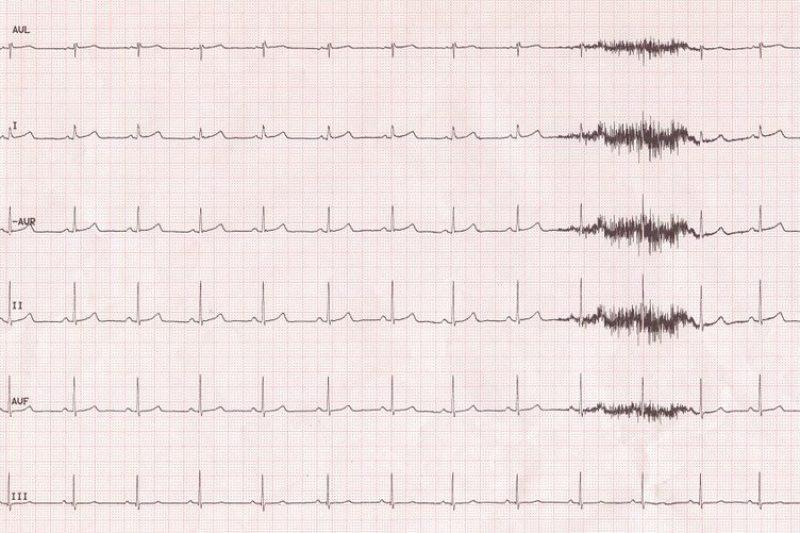
The heart beats due to electrical impulses that travel through specific pathways, prompting each chamber to contract in sequence. An EKG records this activity through small electrodes placed on the chest, arms, and legs. The result is a graph showing the timing and pattern of each heartbeat.
Each wave on the graph corresponds to a specific phase of the heartbeat. If those waves deviate from expected shapes or timing, it can signal a problem with how the heart is functioning.
Conditions that EKGs can detect
While not all heart conditions are visible on an EKG, many common and serious problems show up clearly. Here are several types of heart issues that can often be diagnosed or suggested by an EKG.
Arrhythmias (irregular heart rhythms)
One of the most common reasons for ordering an EKG is to evaluate a possible arrhythmia. These irregular rhythms include:
- Atrial fibrillation (AFib): fast, disorganized beats in the upper chambers
- Bradycardia: abnormally slow heartbeat
- Tachycardia: abnormally fast heartbeat
- Premature beats: extra heartbeats that interrupt the normal rhythm
Each of these shows up as a disruption in the regular spacing or shape of EKG waveforms.
Heart attack (myocardial infarction)
During a heart attack, the blood flow to part of the heart is blocked, causing tissue damage. An EKG can detect these changes in real time, often within minutes of onset. It may show:
- ST segment elevation (indicating active heart attack)
- T wave inversion or Q waves (suggesting past damage)
In emergency rooms, EKGs are one of the first tests done for chest pain or suspected heart attack.
Ischemia (lack of oxygen)
Even if a heart attack hasn’t occurred, the heart may not be getting enough oxygen due to narrowed arteries. EKGs can detect ischemia through subtle waveform changes that indicate reduced blood flow.
Electrolyte imbalances
Abnormal levels of potassium, calcium, or magnesium can change how electrical signals move through the heart. These changes may appear as unusual patterns or wave shapes, alerting doctors to possible metabolic concerns.
Heart enlargement (hypertrophy)
If the heart muscle becomes thickened or stretched—often due to high blood pressure or valve disease—it may generate stronger or altered electrical signals. These appear on the EKG as changes in wave height, direction, or timing.
Conduction blockages
The heart’s electrical signals follow a path through nodes and fibers. If there’s a delay or blockage—such as a bundle branch block or AV block—the EKG will show distinct gaps or changes in wave intervals. These issues may require monitoring or treatment depending on severity.
Pericarditis and other inflammation
When the heart’s outer lining becomes inflamed, as in pericarditis, the EKG may show specific elevations or depressions across multiple leads. These clues help differentiate inflammation from other causes of chest pain.
What EKGs cannot always detect
While EKGs are powerful, they have limitations. Some conditions may not be present during the short recording window or may require further testing to confirm. These include:
- Coronary artery disease without active symptoms
- Valve disorders not affecting rhythm
- Intermittent arrhythmias not captured during the test
In these cases, your doctor may order additional tests like an echocardiogram, Holter monitor, or cardiac stress test to gather more information.
How to interpret EKG findings with your doctor
Most patients will not read their own EKG results. The test is interpreted by a trained healthcare provider or cardiologist who looks at:
- Heart rate and rhythm
- Wave intervals and shapes
- Electrical axis (direction of signal flow)
- Patterns across multiple leads for consistency
The provider will explain whether your EKG is normal or if any findings require further evaluation. Many issues are minor or temporary, but some may point to a need for treatment or additional testing.
When an EKG is recommended
Your provider may suggest an EKG if you are experiencing:
- Chest pain or discomfort
- Shortness of breath
- Rapid or irregular heartbeat
- Fainting or dizziness
- Fatigue without clear cause
It’s also a standard test before some surgeries, during annual exams for high-risk patients, or as a baseline for comparison in future visits.
Even without symptoms, people with a family history of heart disease or risk factors like high blood pressure or diabetes may benefit from occasional EKGs as part of preventive care.
Takeaway
An EKG is a vital diagnostic tool that reveals how your heart is functioning on an electrical level. From arrhythmias to heart attacks and conduction issues, this quick test can uncover a range of conditions that might otherwise go unnoticed. Used wisely, it helps guide early treatment, prevent complications, and support long-term heart health.